数据与计算发展前沿 ›› 2024, Vol. 6 ›› Issue (2): 117-133.
CSTR: 32002.14.jfdc.CN10-1649/TP.2024.02.011
doi: 10.11871/jfdc.issn.2096-742X.2024.02.011
叶旭1( ),杜一1,崔文娟1,沈俊杰2,谢靖2,王露笛1,*()
收稿日期:2023-04-06
出版日期:2024-04-20
发布日期:2024-04-26
通讯作者:
*王露笛(E-mail: 作者简介:叶旭,中国科学院计算机网络信息中心,硕士研究生,主要研究方向为机器学习、数据挖掘等。基金资助:
YE Xu1(),DU Yi1,CUI Wenjuan1,SHEN Junjie2,XIE Jing2,WANG Ludi1,*()
Received:2023-04-06
Online:2024-04-20
Published:2024-04-26
摘要:
【应用背景】随着数据的爆炸式增长、算法的不断改进以及计算能力的快速发展,机器学习在教育、金融、制造和医疗等领域均得到了广泛应用。在眼健康领域,机器学习也已经在疾病诊断、疾病分级、医学检查和疾病早期筛查等许多任务上实现了初步应用。【方法】本文通过对眼健康领域国内外相关文献的调研,从眼科疾病类别、就诊阶段、数据类型及技术类型4个不同维度对领域应用进行了梳理与分析,并对接下来的研究做出相应的展望。【结果】基于调研分析的结果可以看出,在眼健康领域中,机器学习技术主要以图像数据为主,围绕疾病诊断与分级展开。同时在疾病早期筛查和疾病风险预测等处于疾病发展早期阶段的任务上也取得了不错的表现。【结论】通过将机器学习技术应用到眼科诊疗过程的各个阶段,有望降低眼科医生负担、提升眼科医生工作效率、帮助控制患者病情发展、提升患者生活质量并降低患者治疗的经济成本和时间成本。
叶旭, 杜一, 崔文娟, 沈俊杰, 谢靖, 王露笛. 机器学习技术在眼健康领域的应用[J]. 数据与计算发展前沿, 2024, 6(2): 117-133.
YE Xu, DU Yi, CUI Wenjuan, SHEN Junjie, XIE Jing, WANG Ludi. Application of Machine Learning Technology in the Field of Eye Health[J]. Frontiers of Data and Computing, 2024, 6(2): 117-133, https://cstr.cn/32002.14.jfdc.CN10-1649/TP.2024.02.011.
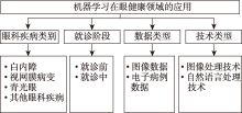
图1
本文综述维度"
表1
从针对的疾病角度对研究的总结"
| 作者 | 年份 | 疾病 | 模型 | 数据 |
|---|---|---|---|---|
| Gao等[ | 2015 | 白内障 | CNN | 裂隙灯图像 |
| Zhang等[ | 2017 | 白内障 | CNN | 眼底图像 |
| Pratap等[ | 2019 | 白内障 | CNN(AlexNet) | 眼底图像 |
| Fraccaro等[ | 2015 | 年龄相关性 黄斑变性 | SVM/随机森 林等 | 电子病历 |
| Lee等[ | 2017 | 年龄相关性 黄斑变性 | CNN | OCT图像 |
| Burlina等[ | 2018 | 年龄相关性 黄斑变性 | CNN | 眼底图像 |
| Oh等[ | 2013 | 糖尿病视网 膜病变 | LASSO等 | 电子健康 记录 |
| 翁铭等[ | 2018 | 糖尿病视网 膜病变 | CNN | 眼底图像 |
| 马菲妍等[ | 2022 | 糖尿病视网 膜病变 | CNN | 眼底图像 |
| Brown等[ | 2018 | 早产儿视网 膜病变 | CNN | 视网膜图像 |
| Gupta等[ | 2019 | 早产儿视网 膜病变 | CNN | 视网膜图像 |
| Thompson等[ | 2020 | 青光眼 | CNN | SD-OCT 图像 |
| Christopher等[ | 2018 | 青光眼 | 多种CNN | 眼底图像 |
| Liu等[ | 2019 | 青光眼 | CNN | 眼底图像 |
| Apostolova等[ | 2017 | 开放性眼球 损伤 | TF-IDF/ SVM等 | 临床文本 记录 |
| Elsawy等[ | 2021 | 角膜疾病 | CNN (VGG-19) | ASOCT 图像 |
表2
从任务所处的阶段对研究的总结"
| 作者 | 年份 | 任务 | 疾病 | 就诊阶段 | 模型 | 数据 |
|---|---|---|---|---|---|---|
| Baxter等[ | 2019 | 疾病干预预测 | 青光眼 | 就诊前 | 随机森林等 | 电子健康记录 |
| Saleh等[ | 2018 | 疾病风险预测 | 糖尿病视网膜病变 | 就诊前 | 模糊随机森林等 | 电子健康记录 |
| Abràmoff等[ | 2018 | 疾病早期筛查 | 糖尿病视网膜病变 | 就诊前 | CNN | 视网膜图像 |
| De Fauwd等[ | 2018 | 疾病诊断 | 多种视网膜疾病 | 就诊中 | 三维U-Net | 三维OCT图像 |
| Ting等[ | 2019 | 疾病诊断 | 白内障 | 就诊中 | CNN(ResNet) | 裂隙灯图像 |
| Burlina 等[ | 2017 | 疾病诊断 | 年龄相关性黄斑变性 | 就诊中 | CNN | 眼底图像 |
| Taylor等[ | 2019 | 疾病分级 | 早产儿视网膜病变 | 就诊中 | CNN | 后视网膜图像 |
| Kanagasingam等[ | 2018 | 疾病分级 | 糖尿病性视网膜病变 | 就诊中 | CNN | 视网膜图像 |
| Li等[ | 2020 | 医学检查 | 视力威胁危险因素 | 就诊中 | CNN (InceptionResnetV2) | OCT图像 |
| Loo等[ | 2022 | 医学检查 | 视盘异常 | 就诊中 | CNN | 眼底图像 |
表3
知名眼科图像数据库"
| 数据库名称 | 疾病种类 | 采集设备 | 是否OA |
|---|---|---|---|
| Messidor-2 | 糖尿病视网膜病变 | TRC-NW6 non-mydriatic fundus camera (Topcon) | 是 |
| DRIVE | 糖尿病视网膜病变 | CR5 non-mydriatic 3CCD camera (Canon) | 是 |
| EyePACS | 糖尿病视网膜病变 | Centervue DRS (Centervue, 意大利), Optovue iCam (Optovue, 美国), Canon CR1/DGi/CR2 (Canon),和Topcon NW (Topcon) | 是 |
| E-ophtha | 糖尿病视网膜病变 | 未知 | 是 |
| ACRIMA | 青光眼 | TRC retina camera (Topcon, 日本) | 是 |
| Corneal Endothelial Cell | 角膜病 | SP-3000 specular microscope (Topcon) | 是 |
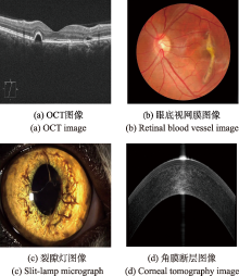
图2
常见眼科图像数据类型"

| [1] | 王坤, 毛阿燕, 孟月莉, 等. 我国公共卫生体系建设发展历程, 现状, 问题与策略[J]. 中国公共卫生, 2019, 35(7): 801-805. |
| [2] | World Health Organization. World report on vision[R]. 2019.Licence:CC BY-NC-SA 3.0 IGO. |
| [3] | Global Health Data Exchange. Global burden of disease study 2019 data resources[EB/OL]. [2020-12-28]. http://ghdx.healthdata.org/gbd-2019. |
| [4] |
CHEN B, JUAN Y E. 中国眼病疾病负担现状及三十年变化趋势[J]. Journal of Zhejiang University (Medical Sciences), 2021, 50(4): 420.
doi: 10.3724/zdxbyxb-2021-0246 |
| [5] | 景正伟, 任贺, 王洪源, 等. 我国眼科资源配置公平性和效率分析[J]. 中国医院管理, 2019, 39(8):36-39. |
| [6] |
NADERI K, GORMLEY J, O’BRART D. Cataract surgery and dry eye disease: a review[J]. European Journal of Ophthalmology, 2020, 30(5): 840-855.
doi: 10.1177/1120672120929958 |
| [7] |
GAO X, LIN S, WONG T Y. Automatic feature learning to grade nuclear cataracts based on deep learning[J]. IEEE Transactions on Biomedical Engineering, 2015, 62(11): 2693-2701.
doi: 10.1109/TBME.2015.2444389 |
| [8] | IAPB Report-State of the World Sight 2010[OL]. Avaliable: http://www.iapb.org/resource/iapb-report-state-world-sight-2010. |
| [9] | ZHANG L, LI J, HAN H, et al. Automatic cataract detection and grading using deep convolutional neural network[C]// 2017 IEEE 14th international conference on networking, sensing and control (ICNSC), IEEE, 2017: 60-65. |
| [10] |
PRATAP T, KOKIL P. Computer-aided diagnosis of cataract using deep transfer learning[J]. Biomedical Signal Processing and Control, 2019, 53: 101533.
doi: 10.1016/j.bspc.2019.04.010 |
| [11] |
AL-ZAMIL W M, YASSIN S A. Recent developments in age-related macular degeneration: a review[J]. Clinical Interventions in Aging, 2017, 12: 1313.
doi: 10.2147/CIA |
| [12] |
FRACCARO P, NICOLO M, BONETTO M, et al. Combining macula clinical signs and patient characteristics for age-related macular degeneration diagnosis: a machine learning approach[J]. BMC Ophthalmol, 2015, 15, (1):1-9.
doi: 10.1186/1471-2415-15-1 |
| [13] |
LEE C S, BAUGHMAN D M, LEE A Y. Deep learning is effective for classifying normal versus age-related macular degeneration OCT images[J]. Ophthalmology Retina, 2017, 1(4): 322-327.
doi: 10.1016/j.oret.2016.12.009 |
| [14] |
BURLINA P M, JOSHI N, PACHECO K D, et al. Use of deep learning for detailed severity characterization and estimation of 5-year risk among patients with age-related macular degeneration[J]. JAMA Ophthalmology, 2018, 136(12): 1359-1366.
doi: 10.1001/jamaophthalmol.2018.4118 pmid: 30242349 |
| [15] | 陈雪珍, 吴慧华, 刘媛媛, 等. 糖尿病视网膜病变患病率的Meta分析[J]. 中国公共卫生管理, 2020, 36(4): 460-465. |
| [16] | SHAH A. Prevalence of diabetic retinopathy in the United States, 2011—2014[J]. Value in Health, 2016, 19(3): A199. |
| [17] | LI Y, TENG D, SHI X, et al. Prevalence of diabetes recorded in mainland China using 2018 diagnostic criteria from the American Diabetes Association: national cross sectional study[J]. British Medical Journal, 2020, 369: 997. |
| [18] |
MANSBERGER S L, SHEPPLER C, BARKER G, et al. Long-term comparative effectiveness of telemedicine in providing diabetic retinopathy screening examinations: a randomized clinical trial[J]. JAMA ophthalmology, 2015, 133(5): 518-525.
doi: 10.1001/jamaophthalmol.2015.1 pmid: 25741666 |
| [19] |
OH E, YOO T K, PARK E C. Diabetic retinopathy risk prediction for fundus examination using sparse learning: a cross-sectional study[J]. BMC Medical Informatics and Decision Making, 2013, 13(1): 1-14.
doi: 10.1186/1472-6947-13-1 |
| [20] |
SALEH E, BŁASZCZYŃSKI J, MORENO A, et al. Learning ensemble classifiers for diabetic retinopathy assessment[J]. Artificial Intelligence in Medicine, 2018, 85: 50-63.
doi: S0933-3657(17)30059-3 pmid: 28993124 |
| [21] | 翁铭, 郑博, 吴茂念, 等. 基于深度学习的DR筛查智能诊断系统的初步研究[J]. 国际眼科杂志, 2018, 18(3): 568-571. |
| [22] | 马菲妍, 张彩霞, 冬雪川. 基于多光谱眼底成像开发的人工智能对糖尿病视网膜病早期体征的识别[J]. 吉林医学, 2022, 43(2): 386-390. |
| [23] |
BROWN J M, CAMPBELL J P, BEERS A, et al. Automated diagnosis of plus disease in retinopathy of prematurity using deep convolutional neural networks[J]. JAMA Ophthalmology, 2018, 136(7): 803-810.
doi: 10.1001/jamaophthalmol.2018.1934 pmid: 29801159 |
| [24] |
GUPTA K, CAMPBELL J P, TAYLOR S, et al. A quantitative severity scale for retinopathy of prematurity using deep learning to monitor disease regression after treatment[J]. JAMA Ophthalmology, 2019, 137(9): 1029-1036.
doi: 10.1001/jamaophthalmol.2019.2442 |
| [25] |
TAYLOR S, BROWN J M, GUPTA K, et al. Monitoring disease progression with a quantitative severity scale for retinopathy of prematurity using deep learning[J]. JAMA Ophthalmology, 2019, 137(9): 1022-1028.
doi: 10.1001/jamaophthalmol.2019.2433 |
| [26] |
THAM Y C, LI X, WONG T Y, et al. Global prevalence of glaucoma and projections of glaucoma burden through 2040: A systematic review and meta-analysis[J]. Ophthalmology, 2014, 121(11): 2081-2090.
doi: 10.1016/j.ophtha.2014.05.013 |
| [27] |
VARMA R, LEE P P, GOLDBERG I, et al. An assessment of the health and economic burdens of glaucoma[J]. American Journal of Ophthalmology, 2011, 152(4): 515-522.
doi: 10.1016/j.ajo.2011.06.004 pmid: 21961848 |
| [28] | KIM S J, CHO K J, OH S. Development of machine learning models for diagnosis of glaucoma[J]. PloS One, 2017, 12(5): e0177726. |
| [29] |
RAN A R, THAM C C, CHAN P P, et al. Deep learning in glaucoma with optical coherence tomography: a review[J]. Eye, 2021, 35(1): 188-201.
doi: 10.1038/s41433-020-01191-5 |
| [30] | MAYA S, MORINO K, YAMANISHI K. Predicting glaucoma progression using multi-task learning with heterogeneous features[C]// 2014 IEEE International Conference on Big Data (Big Data), IEEE, 2014: 261-270. |
| [31] |
THOMPSON A C, JAMMAL A A, BERCHUCK S I, et al. Assessment of a segmentation-free deep learning algorithm for diagnosing glaucoma from optical coherence tomography scans[J]. JAMA Ophthalmology, 2020, 138(4): 333-339.
doi: 10.1001/jamaophthalmol.2019.5983 pmid: 32053142 |
| [32] | CHRISTOPHER M, BELGHITH A, BOWD C, et al. Performance of deep learning architectures and transfer learning for detecting glaucomatous optic neuropathy in fundus photographs[J]. Scientific Reports, 2018, 8(1): 1-13. |
| [33] |
LIU H, LI L, WORMSTONE I M, et al. Development and validation of a deep learning system to detect glaucomatous optic neuropathy using fundus photographs[J]. JAMA Ophthalmology, 2019, 137(12): 1353-1360.
doi: 10.1001/jamaophthalmol.2019.3501 |
| [34] | APOSTOLOVA E, WHITE H A, MORRIS P A, et al. Open globe injury patient identification in warfare clinical notes[C]// AMIA annual symposium proceedings. American Medical Informatics Association, 2017: 403. |
| [35] |
ELSAWY A, ELEIWA T, CHASE C, et al. Multidisease deep learning neural network for the diagnosis of corneal diseases[J]. American Journal of Ophthalmology, 2021, 226: 252-261.
doi: 10.1016/j.ajo.2021.01.018 pmid: 33529589 |
| [36] |
XIE Y, ZHAO L, YANG X, et al. Screening candidates for refractive surgery with corneal tomographic-based deep learning[J]. JAMA Ophthalmology, 2020, 138(5): 519-526.
doi: 10.1001/jamaophthalmol.2020.0507 pmid: 32215587 |
| [37] |
BAXTER S L, MARKS C, KUO T T, et al. Machine learning-based predictive modeling of surgical intervention in glaucoma using systemic data from electronic health records[J]. American Journal of Ophthalmology, 2019, 208: 30-40.
doi: S0002-9394(19)30328-9 pmid: 31323204 |
| [38] |
SALEH E, BŁASZCZYŃSKI J, MORENO A, et al. Learning ensemble classifiers for diabetic retinopathy assessment[J]. Artificial Intelligence in Medicine, 2018, 85: 50-63.
doi: S0933-3657(17)30059-3 pmid: 28993124 |
| [39] |
ABRÀMOFF M D, LAVIN P T, BIRCH M, et al. Pivotal trial of an autonomous AI-based diagnostic system for detection of diabetic retinopathy in primary care offices[J]. NPJ Digital Medicine, 2018, 1(1): 1-8.
doi: 10.1038/s41746-017-0008-y |
| [40] |
DE FAUW J, LEDSAM J R, ROMERA-PAREDES B, et al. Clinically applicable deep learning for diagnosis and referral in retinal disease[J]. Nature Medicine, 2018, 24(9): 1342-1350.
doi: 10.1038/s41591-018-0107-6 pmid: 30104768 |
| [41] |
TOWNSEND K A, WOLLSTEIN G, DANKS D, et al. Heidelberg Retina Tomograph 3 machine learning classifiers for glaucoma detection[J]. British Journal of Ophthalmology, 2008, 92(6): 814-818.
doi: 10.1136/bjo.2007.133074 |
| [42] |
TING D S J, ANG M, MEHTA J S, et al. Artificial intelligence-assisted telemedicine platform for cataract screening and management: a potential model of care for global eye health[J]. British Journal of Ophthalmology, 2019, 103(11): 1537-1538.
doi: 10.1136/bjophthalmol-2019-315025 |
| [43] |
BURLINA P M, JOSHI N, PEKALA M, et al. Automated grading of age-related macular degeneration from color fundus images using deep convolutional neural networks[J]. JAMA Ophthalmology, 2017, 135(11): 1170-1176.
doi: 10.1001/jamaophthalmol.2017.3782 pmid: 28973096 |
| [44] | KANAGASINGAM Y, XIAO D, VIGNARAJAN J, et al. Evaluation of artificial intelligence-based grading of diabetic retinopathy in primary care[J]. JAMA Network Open, 2018, 1(5): e182665-e182665. |
| [45] | LI Y, FENG W, ZHAO X, et al. Development and validation of a deep learning system to screen vision-threatening conditions in high myopia using optical coherence tomography images[J]. British Journal of Ophthalmology, 2020, 10(4): 500-503. |
| [46] |
PHAM T H, DEVALLA S K, ANG A, et al. Deep learning algorithms to isolate and quantify the structures of the anterior segment in optical coherence tomography images[J]. British Journal of Ophthalmology, 2021, 105(9): 1231-1237.
doi: 10.1136/bjophthalmol-2019-315723 |
| [47] |
LOO J, CAI C X, CHOONG J, et al. Deep learning-based classification and segmentation of retinal cavitations on optical coherence tomography images of macular telangiectasia type 2[J]. British Journal of Ophthalmology, 2022, 106(3): 396-402.
doi: 10.1136/bjophthalmol-2020-317131 |
| [48] |
MILEA D, NAJJAR R P, JIANG Z, et al. Artificial intelligence to detect papilledema from ocular fundus photographs[J]. New England Journal of Medicine, 2020, 382(18): 1687-1695.
doi: 10.1056/NEJMoa1917130 |
| [49] | KIHARA Y, HEEREN T F C, LEE C S, et al. Estimating retinal sensitivity using optical coherence tomography with deep-learning algorithms in macular telangiectasia type 2[J]. JAMA Network Open, 2019, 2(2): e188029-e188029. |
| [50] | 中国医药教育协会数字影像与智能医疗专委会, 中国医药教育协会智能医学专委会. 全球眼科图像公开数据库使用指南(2022)[J]. 眼科新进展, 2022, 42(12): 925-932. |
| [51] | ETIENNE, DECENCIÈRE, XI W, et al. Feedback on a publicly distributed image database: the messidor database[J]. Image Analysis & Stereology, 2014, 33(3): 231-234. |
| [52] | STAAL J J, MD ABRAMOFF, NIEMEIJER M, et al. Digital Retinal Image for Vessel Extraction (DRIVE) Database[DB/OL]. [2013]. https://drive.grand.challenge.org. |
| [53] |
GULSHAN V, PENG L, CORAM M, et al. Development and Validation of a Deep Learning Algorithm for Detection of Diabetic Retinopathy in Retinal Fundus Photographs[J]. Jama, 2016, 316(22): 2402-2410.
doi: 10.1001/jama.2016.17216 pmid: 27898976 |
| [54] |
DECENCIERE E, CAZUGUEL G, ZHANG X, et al. TeleOphta: Machine learning and image processing methods for teleophthalmology[J]. IRBM, 2013, 34(2): 196-203.
doi: 10.1016/j.irbm.2013.01.010 |
| [55] |
RIM TH, LEE A Y, TING D S, et al. Detection of features associated with neovascular age-related macular degeneration in ethnically distinct data sets by an optical coherence tomography: trained deep learning algorithm[J]. British Journal of Ophthalmology, 2021, 105(8): 1133-1139.
doi: 10.1136/bjophthalmol-2020-316984 |
| [56] |
KEEL S, WU J, LEE P Y, et al. Visualizing deep learning models for the detection of referable diabetic retinopathy and glaucoma[J]. JAMA Ophthalmology, 2019, 137(3): 288-292.
doi: 10.1001/jamaophthalmol.2018.6035 pmid: 30570648 |
| [57] | 柴文俊. 基于深度学习的眼科疾病智能辅助诊断系统研究与实现[D]. 北京: 北京邮电大学, 2021. |
| [58] | YANG J, YANG Z, MAO Z, et al. Bi-Modal Deep Learning for Recognizing Multiple Retinal Diseases based on Color Fundus Photos and OCT Images[J]. Investigative Ophthalmology & Visual Science, 2021, 62(8): 2107-2107. |
| [59] |
LI X, JIA M, ISLAM M T, et al. Self-supervised feature learning via exploiting multi-modal data for retinal disease diagnosis[J]. IEEE Transactions on Medical Imaging, 2020, 39(12): 4023-4033.
doi: 10.1109/TMI.42 |
| [60] | GASKIN G L, PERSHING S, COLE T S, et al. Predictive modeling of risk factors and complications of cataract surgery[J]. Europe an Journal Ophthalmology, 2016, 26(4): 328-37. |
| [61] |
DANIEL S K, MICHAEL G, WENJIA C, et al. Identifying Medical Diagnoses and Treatable Diseases by Image-Based Deep Learning[J]. Cell, 2018, 172(5): 1122-1131.
doi: S0092-8674(18)30154-5 pmid: 29474911 |
| [62] | 赵京胜, 宋梦雪, 高祥, 等. 自然语言处理中的文本表示研究[J]. 软件学报, 2021, 33(1): 102-128. |
| [63] | ZHANG N, JIA Q, YIN K, et al. Conceptualized representation learning for chinese biomedical text mining[J]. arXiv preprint arXiv: 2008, 10813. |
| [64] | VASWANI A, SHAZEER N, PARMAR N, et al. Attention is all you need[C]// NIPS'17: Proceedings of the 31st International Conference on Neural Information Processing Systems ACM, 2017: 6000-6010. |
| [65] | DEVLIN J, CHANG M W, LEE K, et al. Bert: Pre-training of deep bidirectional transformers for language understanding[J]. arXiv preprint arXiv: 1810. 04805, 2018. |
| [66] | YANG Z, DAI Z, YANG Y, et al. XLNet: generalized autoregressive pretraining for language understanding[C]// Proceedings of the 33rd International Conference on Neural Information Processing Systems, 2019: 5753-5763. |
| [67] | LIU Y, OTT M, GOYAL N, et al. Roberta: A robustly optimized bert pretraining approach[J]. arXiv preprint arXiv: 1907.11692, 2019. |
| [68] | ALSENTZER E, MURPHY J R, BOAG W, et al. Publicly available clinical BERT embeddings[J]. arXiv preprint arXiv: 1904.03323, 2019. |
| [69] |
LEE J, YOON W, KIM S, et al. BioBERT: a pre-trained biomedical language representation model for biomedical text mining[J]. Bioinformatics, 2020, 36(4): 1234-1240.
doi: 10.1093/bioinformatics/btz682 pmid: 31501885 |
| [70] | SUN Y, WANG S, LI Y, et al. Ernie 2.0: A continual pre-training framework for language understanding[C]// Proceedings of the AAAI Conference on Artificial Intelligence, 2020, 34(5): 8968-8975. |
| [71] | ZHANG N, JIA Q, YIN K, et al. Conceptualized representation learning for chinese biomedical text mining[J]. arXiv preprint arXiv:2008.10813, 2020. |
| [72] | XIAO M, QIAO Z, FU Y, et al. Hierarchical interdisciplinary topic detection model for research proposal classification[J/OL]. IEEE Transactions on Knowledge and Data Engineering, 2023, https://www.doi.org/10.1109/TKDE.2023.3248608. |
| [73] | YE X, XIAO M, NING Z, et al. NEEDED: Introducing Hierarchical Transformer to Eye Diseases Diagnosis[C]// Proceedings of the 2023 SIAM International Conference on Data Mining (SDM), Society for Industrial and Applied Mathematics, 2023: 667-675. |
| [74] | XIAO M, QIAO Z, FU Y, et al. Expert knowledge-guided length-variant hierarchical label generation for proposal classification[C]// 2021 IEEE International Conference on Data Mining (ICDM), IEEE, 2021: 757-766. |
| [75] | MARINHO Z, MENDES A, MIRANDA S, et al. Hierarchical nested named entity recognition[C]// Proceedings of the 2nd Clinical Natural Language Processing Workshop, 2019: 28-34. |
| [76] | JI Z, XIA T, HAN M, et al. A Neural Transition-based Joint Model for Disease Named Entity Recognition and Normalization[C]// Proceedings of the 59th Annual Meeting of the Association for Computational Linguistics and the 11th International Joint Conference on Natural Language Processing (Volume 1:Long Papers), 2021: 2819-2827. |
| [77] | ZHAO S, LIU T, ZHAO S, et al. A neural multi-task learning framework to jointly model medical named entity recognition and normalization[C]// Proceedings of the AAAI Conference on Artificial Intelligence, 2019, 33(1): 817-824. |
| [78] | CHEN M, LAN G, DU F, et al. Joint Learning with Pre-trained Transformer on Named Entity Recognition and Relation Extraction Tasks for Clinical Analytics[C]// Proceedings of the 3rd Clinical Natural Language Processing Workshop, 2020: 234-242. |
| [79] | SAKAKINI T, LEE J Y, DURI A, et al. Context-Aware Automatic Text Simplification of Health Materials in Low-Resource Domains[C]// Proceedings of the 11th International Workshop on Health Text Mining and Information Analysis, 2020: 115-126. |
| [80] | LIANG J, TSOU C H, PODDAR A. A novel system for extractive clinical note summarization using EHR data[C]// Proceedings of the 2nd clinical natural language processing workshop, 2019: 46-54. |
| [81] | CHEN J, DAI X, YUAN Q, et al. Towards interpretable clinical diagnosis with bayesian network ensembles stacked on entity-aware cnns[C]// Proceedings of the 58th Annual Meeting of the Association for Computational Linguistics, 2020: 3143-3153. |
| [82] | YUAN Q, CHEN J, LU C, et al. The graph-based mutual attentive network for automatic diagnosis[C]// Proceedings of the Twenty-Ninth International Conference on International Joint Conferences on Artificial Intelligence, 2021: 3393-3399. |
| [83] | YANG Z, HUANG Y, JIANG Y, et al. Clinical assistant diagnosis for electronic medical record based on convolutional neural network[J]. Scientific Reports, 2018, 8(1): 1-9. |
| [84] | YUWONO S K, NG H T, NGIAM K Y. Learning from the experience of doctors: automated diagnosis of appendicitis based on clinical notes[C]// Proceedings of the 18th BioNLP Workshop and Shared Task, 2019: 11-19. |
| [85] | FENG J, SHAIB C, RUDZICZ F. Explainable clinical decision support from text[C]// Proceedings of the 2020 Conference on Empirical Methods in Natural Language Processing (EMNLP), 2020: 1478-1489. |
| [86] | SI Y, ROBERTS K. Hierarchical Transformer Networks for Longitudinal Clinical Document Classification[J]. arXiv preprint arXiv:2104.08444, 2021. |
| [87] |
HASHIR M, SAWHNEY R. Towards unstructured mortality prediction with free-text clinical notes[J]. Journal of Biomedical Informatics, 2020, 108: 103489.
doi: 10.1016/j.jbi.2020.103489 |
| [88] | MULLENBACH J, WIEGREFFE S, DUKE J, et al. Explainable prediction of medical codes from clinical text[J]. arXiv preprint arXiv:1802.05695, 2018. |
| [89] | ZHANG Z, LIU J, RAZAVIAN N. BERT-XML: Large scale automated ICD coding using BERT pretraining[J]. arXiv preprint arXiv:2006.03685, 2020. |
| [1] | 何睿琳, 杨欣怡, 孙洪赞, 李晨. 基于图特征的组织病理学图像分析方法的最新发展情况与展望[J]. 数据与计算发展前沿, 2024, 6(2): 101-116. |
| [2] | 申志豪, 李娜, 尹世豪, 杜一, 胡良霖. 基于TPA-Transformer的机票价格预测[J]. 数据与计算发展前沿, 2023, 5(6): 115-125. |
| [3] | 危婷, 彭亮, 牛铁, 张宏海. 基于特征分析的HPC失败作业的检测和根因分析[J]. 数据与计算发展前沿, 2023, 5(6): 94-103. |
| [4] | 孙一帆, 张锐, 陶杨, 高碧柔, 秦诗涵, 安超. 本地化差分隐私综述[J]. 数据与计算发展前沿, 2023, 5(5): 74-97. |
| [5] | 田一擎, 程曦, 冯博靖. 企业信用评级计算模型综述[J]. 数据与计算发展前沿, 2023, 5(4): 139-153. |
| [6] | 陈美霖, 刘端阳, 徐黎明, 汪洋. 基于机器学习的力场模型研究综述[J]. 数据与计算发展前沿, 2023, 5(4): 27-37. |
| [7] | 刘端阳, 魏钟鸣. 有监督学习算法在材料科学中的应用[J]. 数据与计算发展前沿, 2023, 5(4): 38-47. |
| [8] | 李妍,何洪波,王闰强. 微博热度预测研究综述[J]. 数据与计算发展前沿, 2023, 5(2): 119-135. |
| [9] | 高添,朱教君,张金鑫,孙一荣,于丰源,滕德雄,卢德亮,于立忠,王宗国. 基于新一代信息技术的温带森林生态系统碳通量精准计量[J]. 数据与计算发展前沿, 2023, 5(2): 60-72. |
| [10] | 王凡,冯立强,曹荣强. 大数据驱动的海洋人工智能服务平台设计与应用[J]. 数据与计算发展前沿, 2023, 5(2): 73-85. |
| [11] | 赵忠斌,蔡满春,芦天亮. 融合多头注意力机制的网络恶意流量检测[J]. 数据与计算发展前沿, 2022, 4(5): 60-67. |
| [12] | 危婷,张宏海,蔺小丽,张蕾蕾,王妍,贾金峰. 云服务网站用户复访行为预测模型研究[J]. 数据与计算发展前沿, 2022, 4(3): 124-130. |
| [13] | 孙永谦,张茹茹,林子涵,张圣林,谭智元,张玉志. KPI异常检测方法评估[J]. 数据与计算发展前沿, 2022, 4(3): 46-65. |
| [14] | 张怡宁,何洪波,王闰强. 热门数字音频预测技术综述[J]. 数据与计算发展前沿, 2021, 3(4): 81-92. |
| [15] | 蒲剑苏,朱正国,邵慧,高博洋,朱焱麟,闫宗楷,向勇. 基于可视化的固态电解质材料机器学习筛选与预测[J]. 数据与计算发展前沿, 2021, 3(4): 18-29. |
| 阅读次数 | ||||||
|
全文 |
|
|||||
|
摘要 |
|
|||||