数据与计算发展前沿 ›› 2024, Vol. 6 ›› Issue (3): 3-14.
CSTR: 32002.14.jfdc.CN10-1649/TP.2024.03.001
doi: 10.11871/jfdc.issn.2096-742X.2024.03.001
蔡程飞1,3( ),李军2,焦一平2,王向学2,郭冠辰1,徐军2,*()
收稿日期:2023-10-21
出版日期:2024-06-20
发布日期:2024-06-21
通讯作者:
*徐军(E-mail: 作者简介:蔡程飞,南京信息工程大学自动化学院,博士研究生,主要研究方向为病理图像计算、多模态信息融合。基金资助:
CAI Chengfei1,3(),LI Jun2,JIAO Yiping2,WANG Xiangxue2,GUO Guanchen1,XU Jun2,*()
Received:2023-10-21
Online:2024-06-20
Published:2024-06-21
摘要:
【目的】在肿瘤学中,患者有一系列的临床数据,从放射学、组织学、基因组学到电子健康记录。不同数据模式的整合为提高诊断和预后模型的稳健性和准确性提供了机会,使人工智能在临床实践发挥重要作用。【方法】本文将探讨深度学习技术以及其在肿瘤医学数据中的应用,并研究肿瘤学领域多模态数据融合方法的潜在影响和重要发现。【结果】多模态数据能够更好地发现与患者治疗响应、预后效果相关的信息,从而构建更加鲁棒的深度学习模型。【结论】深度学习已经在医学领域取得了显著的进展,特别是在肿瘤学研究中处理多模态医学数据。这些进展为临床提供了更准确、更快速的工具来进行肿瘤的检测、分割、分类和预后预测,同时也面临很多挑战亟须解决。
蔡程飞, 李军, 焦一平, 王向学, 郭冠辰, 徐军. 基于深度学习的医学多模态数据融合方法在肿瘤学中的进展和挑战[J]. 数据与计算发展前沿, 2024, 6(3): 3-14.
CAI Chengfei, LI Jun, JIAO Yiping, WANG Xiangxue, GUO Guanchen, XU Jun. Progress and Challenges of Medical Multimodal Data Fusion Methods Based on Deep Learning in Oncology[J]. Frontiers of Data and Computing, 2024, 6(3): 3-14, https://cstr.cn/32002.14.jfdc.CN10-1649/TP.2024.03.001.
图1
整合来自不同数据源的信息和临床背景的整体框架,展开不同的预测任务,助力个体化精准医疗"
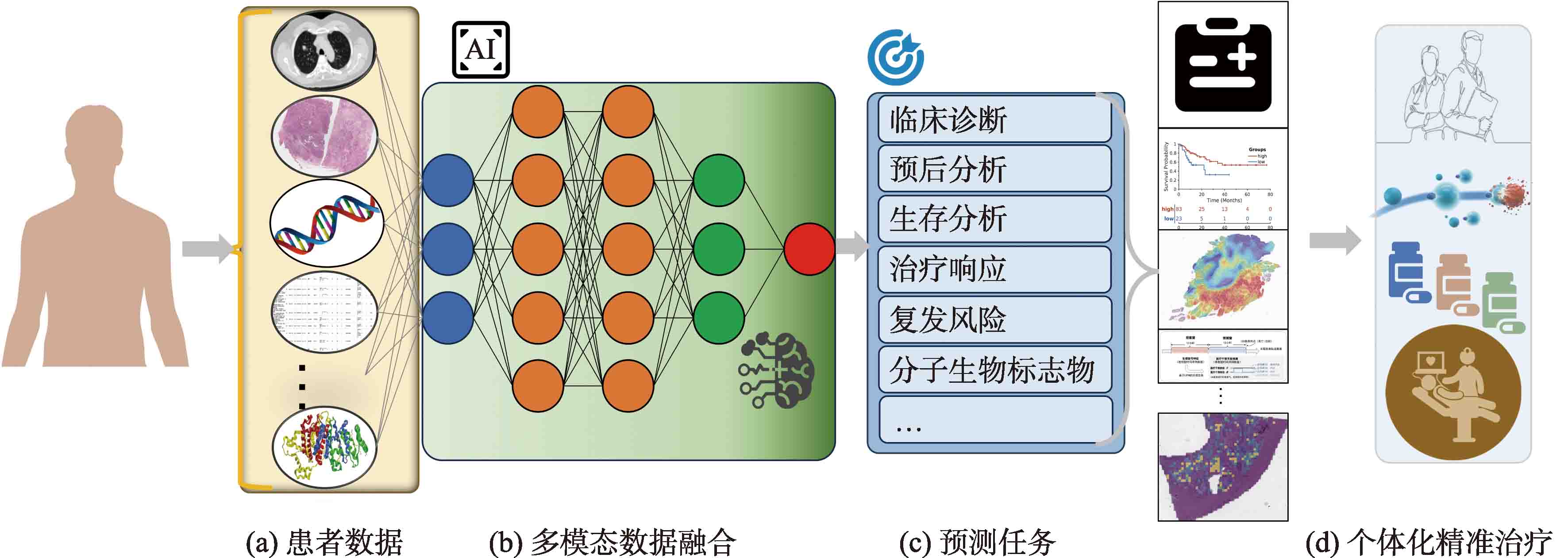

图2
以病理数据为例实现不同类型的算法流程"
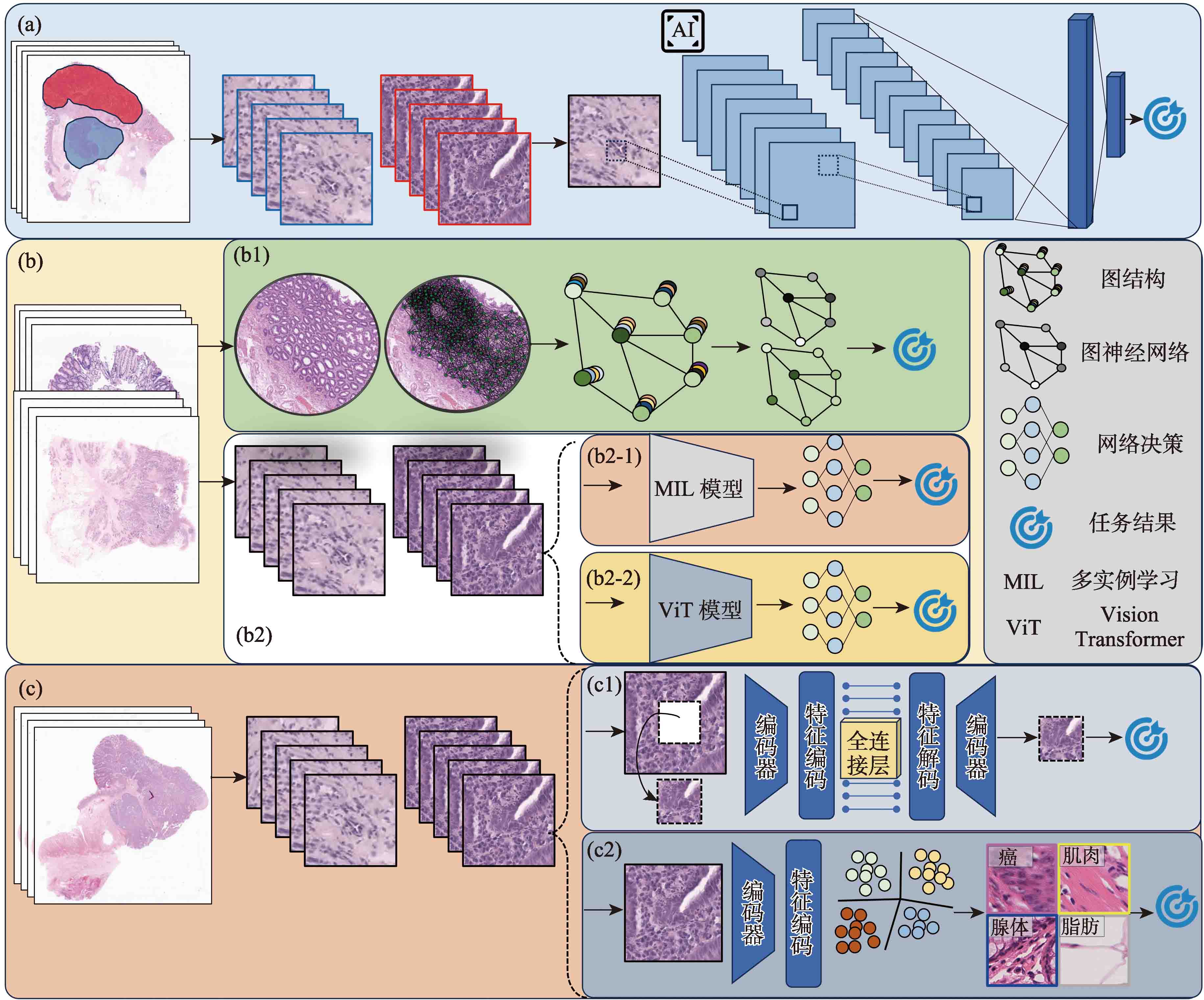
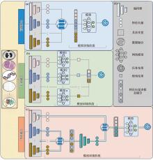
图3
展示多模态数据不同方式的融合流程"
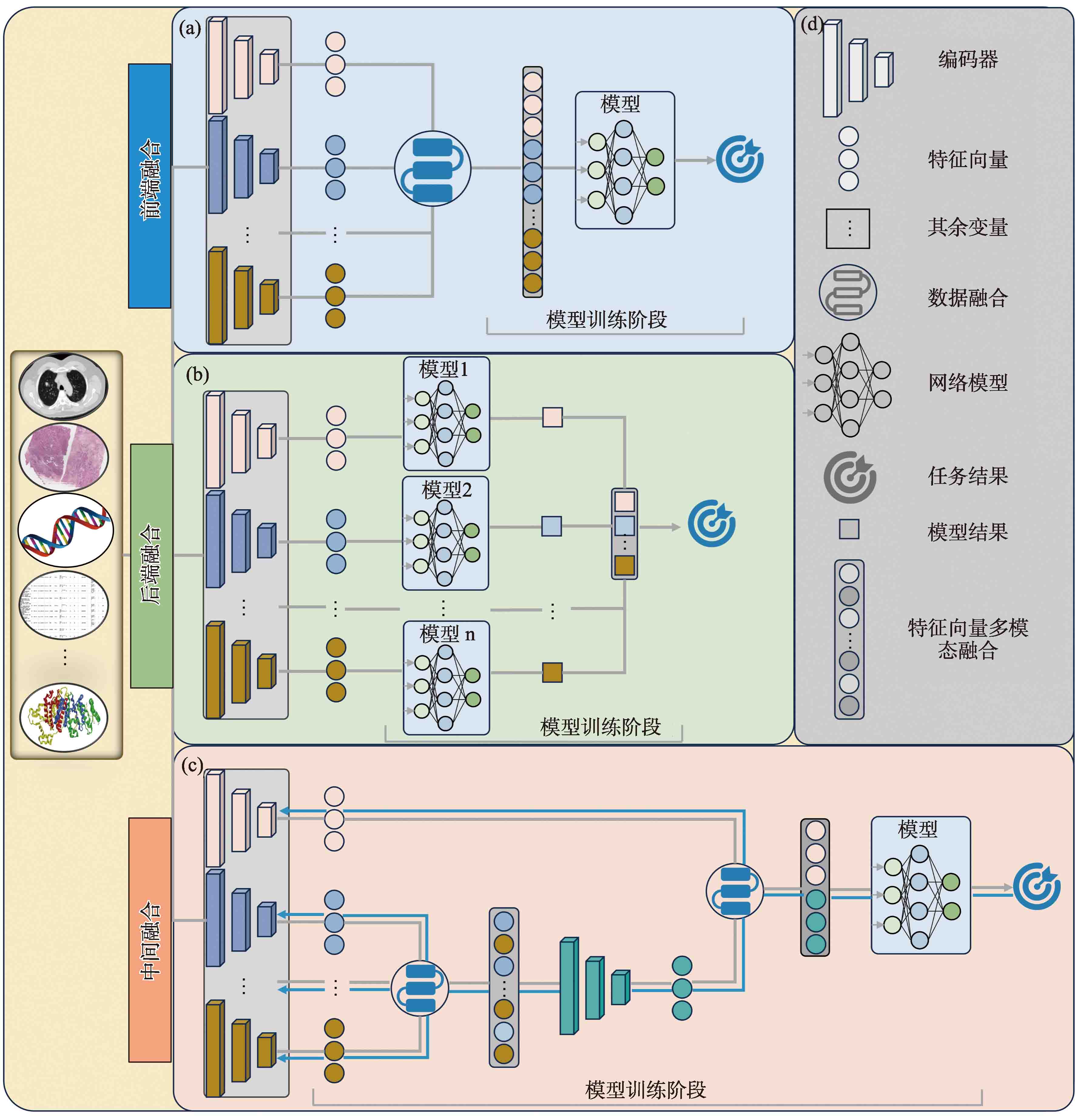
图4
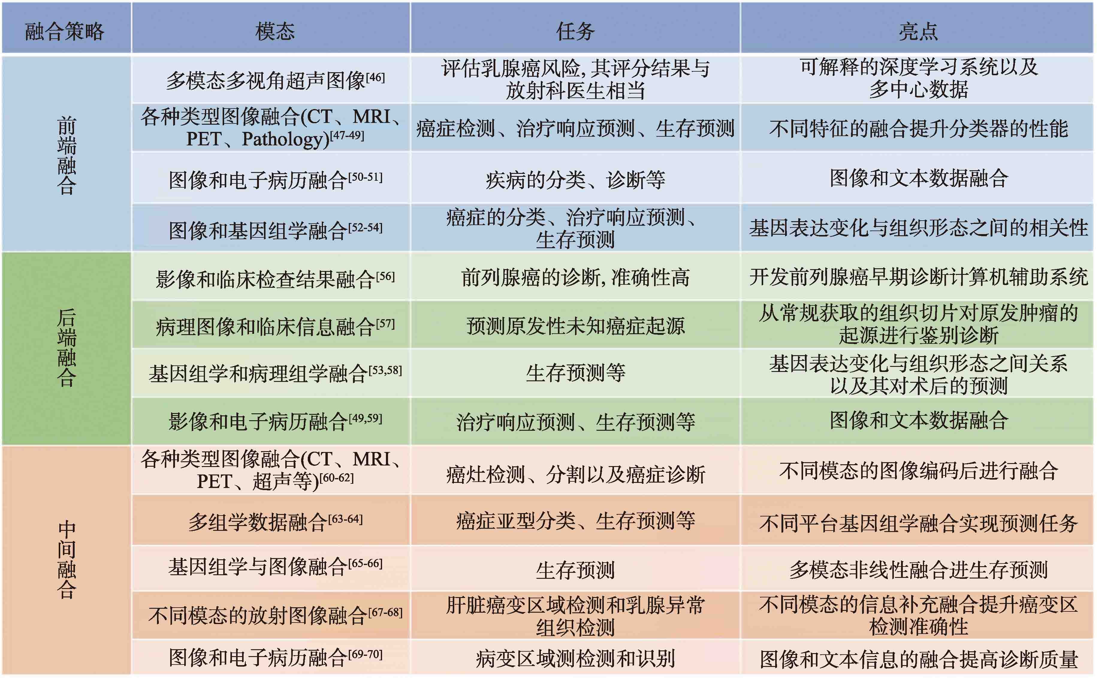
融合策略的总结"
| [1] | GUO Z, et al. Medical image segmentation based on multi-modal convolutional neural network: Study on image fusion schemes[C]. 2018 IEEE 15th International Symposium on Biomedical Imaging, IEEE, 2018: 903-907. |
| [2] |
LI L, et al. Deep learning for variational multimodality tumor segmentation in PET/CT[J]. Neurocomputing, 2020, 392: 277-295.
doi: 10.1016/j.neucom.2018.10.099 pmid: 32773965 |
| [3] | SAEED N, et al. An ensemble approach for patient prognosis of head and neck tumor using multimodal data[J]. 3D Head and Neck Tumor Segmentation in PET/CT Challenge, Cham: Springer International Publishing, 2021, 13209: 278-286. |
| [4] | SHOEIBI A, et al. Diagnosis of brain diseases in fusion of neuroimaging modalities using deep learning: A review[J]. Information Fusion, 2022, 93: 85-117. |
| [5] | BERA K, et al. Artificial intelligence in digital pathology—new tools for diagnosis and precision oncology[J]. Nature reviews Clinical oncology, 2019, 16(11): 703-715. |
| [6] | VU Q D, et al. Handcrafted Histological Transformer (H2T): Unsupervised representation of whole slide images[J]. Medical Image Analysis, 2023. 85: 102743-102761. |
| [7] | HUANG Y, et al. Deep learning radiopathomics based on preoperative US images and biopsy whole slide images can distinguish between luminal and non-luminal tumors in early-stage breast cancers[J]. EBioMedicine, 2023, 94: 104706-104719. |
| [8] | BILAL M, et al. Development and validation of a weakly supervised deep learning framework to predict the status of molecular pathways and key mutations in colorectal cancer from routine histology images: a retrospective study[J]. The Lancet Digital Health, 2021, 3(12): 763-772. |
| [9] | YAMASHITA R, et al. Deep learning model for the prediction of microsatellite instability in colorectal cancer: a diagnostic study[J]. The Lancet Oncology, 2021, 22(1): 132-141. |
| [10] | CHANG X, et al. Predicting colorectal cancer microsatellite instability with a self-attention-enabled convolutional neural network[J]. Cell Reports Medicine, 2023, 4(2): 100914-100930. |
| [11] |
MARCUS L, et al. FDA approval summary: pembrolizumab for the treatment of microsatellite instability-high solid tumors[J]. Clinical Cancer Research, 2019, 25(13): 3753-3758.
doi: 10.1158/1078-0432.CCR-18-4070 pmid: 30787022 |
| [12] | LIU J, et al. Immune landscape and prognostic immune-related genes in KRAS-mutant colorectal cancer patients[J]. Journal of translational medicine, 2021, 19: 1-17. |
| [13] |
WAGNER S J, et al. Transformer-based biomarker prediction from colorectal cancer histology: A large-scale multicentric study[J]. Cancer Cell, 2023, 41(9): 1650-1661.
doi: 10.1016/j.ccell.2023.08.002 pmid: 37652006 |
| [14] | PAI R K, et al. Quantitative pathologic analysis of digitized images of colorectal carcinoma improves prediction of recurrence-free survival[J]. Gastroenterology, 2022, 163(6): 1531-1546. |
| [15] | LI Z, et al. Survival Prediction via Hierarchical Multimodal Co-Attention Transformer: A Computational Histology-Radiology Solution[J]. IEEE Transactions on Medical Imaging, 2023, 42(9): 2678-2689. |
| [16] | BOEHM K.M., et al. Harnessing multimodal data integration to advance precision oncology[J]. Nature Reviews Cancer, 2022, 22(2): 114-126. |
| [17] | RAZZAGHI P, et al. Multimodal brain tumor detection using multimodal deep transfer learning[J]. Applied Soft Computing, 2022, 129: 109631-109642. |
| [18] | XU L, et al. Bi-MGAN: Bidirectional T1-to-T2 MRI images prediction using multi-generative multi-adversarial nets[J]. Biomedical Signal Processing and Control, 2022, 78: 103994-104005. |
| [19] |
HAVAEI M, et al. Brain tumor segmentation with deep neural networks[J]. Medical image analysis, 2017, 35: 18-31.
doi: S1361-8415(16)30033-0 pmid: 27310171 |
| [20] | LI Y, et al. Deep learning based multimodal brain tumor diagnosis[C]. Springer International Publishing, 2018, Revised Selected Papers 3: 149-158. |
| [21] | GAO, ZEYU, et al. A semi-supervised multi-task learning framework for cancer classification with weak annotation in whole-slide images[J]. Medical Image Analysis, 2023, 83: 102652-102666. |
| [22] | GAO, ZEYU, et al. Uncertainty-based Model Acceleration for Cancer Classification in Whole-Slide Images[C]. 2022 IEEE International Conference on Bioinformatics and Biomedicine (BIBM), IEEE, 2022, 1534-1538. |
| [23] | GRAHAM S, et al. Screening of normal endoscopic large bowel biopsies with artificial intelligence: a retrospective study[J]. Gut, 2023, 72: 1709-1721. |
| [24] | ZHANG S, et al. Graph convolutional networks: a comprehensive review[J]. Computational Social Networks, 2019, 6(1): 1-23. |
| [25] | QU L, et al. Towards label-efficient automatic diagnosis and analysis: a comprehensive survey of advanced deep learning-based weakly-supervised, semi-supervised and self-supervised techniques in histopathological image analysis[J]. Physics in Medicine and Biology, 2022, 67(20): 1-35. |
| [26] | FENG J and ZHOU Z. Deep miml network[C]. In Proceedings of the AAAI Conference on Artificial Intelligence (AAAI), 2017, 31(1): 10890-10897. |
| [27] | WANG X, et al. Revisiting multiple instance neural networks[J]. Pattern Recognition, 2018, 74: 15-24. |
| [28] | WU J, et al. Deep multiple instance learning for image classification and auto-annotation[C]. In Proceedings of the IEEE Conference on Computer Vision and Pattern Recognition (CVPR), 2015: 3460-3469. |
| [29] | CHEN T, et al. A simple framework for contrastive learning of visual representations[C]. In International Conference on Machine Learning (ICML), 2020, 1597-1607. |
| [30] | KATHRER J N, et al. Multi-class texture analysis in colorectal cancer histology[J]. Scientific Reports, 2016, 6(1): 1-11. |
| [31] | HASHIMOTO N, et al. Multi-scale domain-adversarial multiple-instance cnn for cancer subtype classification with unannotated histopathological images[C]. In Proceedings of the IEEE/CVF Conference on Computer Vision and Pattern Recognition (CVPR), 2020: 3852-3861. |
| [32] | LI H, et al. Dt-mil: Deformable transformer for multi-instance learning on histopathological image[C]. In International Conference on Medical Image Computing and Computer-Assisted Intervention (MICCAI), Springer, 2021: 206-216. |
| [33] | YAO J, et al. Whole slide images based cancer survival prediction using attention guided deep multiple instance learning networks[J]. Medical Image Analysis, 2020, 65: 101789-101804. |
| [34] | ZHU X, et al. Wsisa: Making survival prediction from whole slide histopathological images[C]. In Proceedings of the IEEE Conference on Computer Vision and Pattern Recognition (CVPR), 2017: 7234-7242. |
| [35] |
CAMPANELLA G, et al. Clinical-grade computational pathology using weakly supervised deep learning on whole slide images[J]. Nature medicine, 2019, 25(8): 1301-1309.
doi: 10.1038/s41591-019-0508-1 pmid: 31308507 |
| [36] |
LU M Y, et al. Data-efficient and weakly supervised computational pathology on whole-slide images[J]. Nature biomedical engineering, 2021, 5(6): 555-570.
doi: 10.1038/s41551-020-00682-w pmid: 33649564 |
| [37] | ILSE M, et al. Attention-based deep multiple instance learning[C]. International conference on machine learning, PMLR, 2018: 2127-2136. |
| [38] | DOSOVITSKIY A, et al. An image is worth 16x16 words: Transformers for image recognition at scale[J]. arXiv preprint, arXiv, 2020, 2010: 11929-11951. |
| [39] | VASWANI A, et al. Attention is all you need[C]. Advances in neural information processing systems, 2017: 30-41. |
| [40] | HU, ZEBIN, et al. Data-enabled intelligence in complex industrial systems cross-model transformer method for medical image synthesis[J]. Complexity, 2021, (2021): 1-7. |
| [41] | TRAGAKIS A, et al. The fully convolutional transformer for medical image segmentation[C]. Proceedings of the IEEE/CVF Winter Conference on Applications of Computer Vision, 2023: 3660-3669. |
| [42] | JING L, et al. Self-supervised visual feature learning with deep neural networks: A survey[J]. IEEE transactions on pattern analysis and machine intelligence, 2020, 43(11): 4037-4058. |
| [43] | KAZEROUNI, et al. Diffusion models for medical image analysis: A comprehensive survey[J]. arXiv preprint arXiv:2211.07804 (2022). |
| [44] | SLEEMANIV W C, et al. Multimodal classification: Current landscape, taxonomy and future directions[J]. ACM Computing Surveys, 2022, 55(7): 1-31. |
| [45] | BALTRUSAITIS T, et al. Multimodal machine learning: A survey and taxonomy[J]. IEEE transactions on pattern analysis and machine intelligence, 2018, 41(2): 423-443. |
| [46] |
QIAN X, et al. Prospective assessment of breast cancer risk from multimodal multiview ultrasound images via clinically applicable deep learning[J]. Nature biomedical engineering, 2021, 5(6): 522-532.
doi: 10.1038/s41551-021-00711-2 pmid: 33875840 |
| [47] |
LE M.H., et al. Automated diagnosis of prostate cancer in multi-parametric MRI based on multimodal convolutional neural networks[J]. Physics in Medicine and Biology, 2017, 62(16): 6497-6514.
doi: 10.1088/1361-6560/aa7731 pmid: 28582269 |
| [48] |
LIPKOVA J, et al. Personalized radiotherapy design for glioblastoma: Integrating mathematical tumor models, multimodal scans, and bayesian inference[J]. IEEE transactions on medical imaging, 2019, 38(8): 1875-1884.
doi: 10.1109/TMI.2019.2902044 pmid: 30835219 |
| [49] |
NIE D, et al. Multi-channel 3D deep feature learning for survival time prediction of brain tumor patients using multi-modal neuroimages[J]. Scientific reports, 2019, 9(1): 1103-1117.
doi: 10.1038/s41598-018-37387-9 pmid: 30705340 |
| [50] |
YAP J, et al. Multimodal skin lesion classification using deep learning[J]. Experimental dermatology, 2018, 27(11): 1261-1267.
doi: 10.1111/exd.13777 pmid: 30187575 |
| [51] | XU T, et al. Multimodal deep learning for cervical dysplasia diagnosis[C]. Medical Image Computing and Computer-Assisted Intervention-MICCAI 2016: 19th, Springer International Publishing, 2016, Proceedings, Part II 19: 115-123. |
| [52] |
KHOSRAVI P, et al. A deep learning approach to diagnostic classification of prostate cancer using pathology-radiology fusion[J]. Journal of Magnetic Resonance Imaging, 2021, 54(2): 462-471.
doi: 10.1002/jmri.27599 pmid: 33719168 |
| [53] |
CHEN R J, et al. Pan-cancer integrative histology-genomic analysis via multimodal deep learning[J]. Cancer Cell, 2022, 40(8): 865-878.
doi: 10.1016/j.ccell.2022.07.004 pmid: 35944502 |
| [54] | SAMMUT S.J., et al. Multi-omic machine learning predictor of breast cancer therapy response[J]. Nature, 2022, 601(7894): 623-629. |
| [55] | RAMANATHAN T T, et al. Naïve Bayes Based Multiple Parallel Fuzzy Reasoning Method for Medical Diagnosis[J]. Journal of Engineering Science and Technology, 2022, 17(1): 472-490. |
| [56] | REDA I, et al. Deep learning role in early diagnosis of prostate cancer[J]. Technology in cancer research and treatment, 2018, 17: 1-11. |
| [57] | LU M Y, et al. AI-based pathology predicts origins for cancers of unknown primary[J]. Nature, 2021, 594(7861): 106-110. |
| [58] | SHA X, et al. Identifying pathological subtypes of non-small-cell lung cancer by using the radiomic features of 18F-fluorodeoxyglucose positron emission computed tomography[J]. Translational Cancer Research, 2019, 8(5): 1741-1749. |
| [59] |
JOO S, et al. Multimodal deep learning models for the prediction of pathologic response to neoadjuvant chemotherapy in breast cancer[J]. Scientific reports, 2021, 11(1): 18800-18808.
doi: 10.1038/s41598-021-98408-8 pmid: 34552163 |
| [60] | KUMAR A, et al. Co-learning feature fusion maps from PET-CT images of lung cancer[J]. IEEE Transactions on Medical Imaging, 2019, 39(1): 204-217. |
| [61] |
SEDGHI A, et al. Improving detection of prostate cancer foci via information fusion of MRI and temporal enhanced ultrasound[J]. International journal of computer assisted radiology and surgery, 2020, 15: 1215-1223.
doi: 10.1007/s11548-020-02172-5 pmid: 32372384 |
| [62] | HAVAEI M, et al. Hemis: Hetero-modal image segmentation[C]. Medical Image Computing and Computer-Assisted Intervention-MICCAI 2016: 19th, Springer International Publishing, 2016, Proceedings, Part II 19: 469-477. |
| [63] | LIANG M, et al. Integrative data analysis of multi-platform cancer data with a multimodal deep learning approach[J]. IEEE/ACM transactions on computational biology and bioinformatics, 2014, 12(4): 928-937. |
| [64] | LAI Y H, et al. Overall survival prediction of non-small cell lung cancer by integrating microarray and clinical data with deep learning[J]. Scientific reports, 2020, 10(1): 4679-4690. |
| [65] | VALE-SILVA L A, et al. Long-term cancer survival prediction using multimodal deep learning[J]. Scientific Reports, 2021, 11(1): 13505-13516. |
| [66] |
YALA A, et al. A deep learning mammography-based model for improved breast cancer risk prediction[J]. Radiology, 2019, 292(1): 60-66.
doi: 10.1148/radiol.2019182716 pmid: 31063083 |
| [67] | MO S, et al. Multimodal priors guided segmentation of liver lesions in MRI using mutual information based graph co-attention networks[C]. Medical Image Computing and Computer Assisted Intervention-MICCAI 2020: 23rd, Springer International Publishing, 2020, Proceedings, Part IV 23:429-438. |
| [68] | LEI B, et al. Self-co-attention neural network for anatomy segmentation in whole breast ultrasound[J]. Medical image analysis, 2020, 64: 101753-101769. |
| [69] | ZHOU L and YAN L. Deep features fusion with mutual attention transformer for skin lesion diagnosis[C]. 2021 IEEE International Conference on Image Processing (ICIP), IEEE, 2021: 3797-3801. |
| [70] | VO H Q, et al. Multimodal Breast Lesion Classification Using Cross-Attention Deep Networks[C]. 2021 IEEE EMBS International Conference on Biomedical and Health Informatics (BHI), IEEE, 2021: 1-4. |
| [1] | 廖立波, 王书栋, 宋维民, 张兆领, 李刚, 黄永盛. CEPC上基于DeepSets模型的喷注标记算法研究[J]. 数据与计算发展前沿, 2024, 6(3): 108-115. |
| [2] | 严瑾, 董科军, 李洪涛. 融合语义和共现特征的Web跟踪器深度识别方法[J]. 数据与计算发展前沿, 2024, 6(3): 127-138. |
| [3] | 王志永, 刘晶晶, 王新明, 陈博文, 聂伟, 张瀚林, 刘洪海. 孤独症人工智能诊疗进展及前沿[J]. 数据与计算发展前沿, 2024, 6(3): 15-27. |
| [4] | 寇大治. 基于深度学习的口腔全景片牙齿自动分割方法[J]. 数据与计算发展前沿, 2024, 6(3): 162-172. |
| [5] | 郑懿诺, 孙沐毅, 张虹云, 张婧, 邓天政, 刘倩. 深度学习在口腔种植影像学中的应用:研究进展与挑战[J]. 数据与计算发展前沿, 2024, 6(3): 41-49. |
| [6] | 袁家琳, 欧阳汝珊, 戴懿, 赖小慧, 马捷, 龚静山. 基于深度学习乳腺X线摄影钙化识别分类模型的临床应用价值[J]. 数据与计算发展前沿, 2024, 6(2): 68-79. |
| [7] | 王子元, 王国中. 改进的轻量级YOLOv5算法在行人检测的应用[J]. 数据与计算发展前沿, 2023, 5(6): 161-172. |
| [8] | 巨家骥, 黄勃, 张帅, 郭茹燕. 融合情感词典和自注意力的双通道情感分析模型[J]. 数据与计算发展前沿, 2023, 5(4): 101-111. |
| [9] | 李俊飞, 徐黎明, 汪洋, 魏鑫. 基于深度学习技术的科技文献引文分类研究综述[J]. 数据与计算发展前沿, 2023, 5(4): 86-100. |
| [10] | 李妍,何洪波,王闰强. 微博热度预测研究综述[J]. 数据与计算发展前沿, 2023, 5(2): 119-135. |
| [11] | 刘云帆,李琦,孙哲南,谭铁牛. 基于生成对抗网络的人脸年龄编辑方法综述[J]. 数据与计算发展前沿, 2023, 5(2): 2-23. |
| [12] | 涂又友,郑奇靖,赵瑾. 基于深度学习方法研究分子/固体界面量子化质子耦合的电荷转移过程[J]. 数据与计算发展前沿, 2023, 5(2): 37-49. |
| [13] | 许淞源,刘峰. ESDRec:一种面向地球大数据平台的数据推荐模型[J]. 数据与计算发展前沿, 2023, 5(1): 55-64. |
| [14] | 刘琦玮,李俊,顾蓓蓓,赵泽方. TSAIE:图像增强文本的多模态情感分析模型[J]. 数据与计算发展前沿, 2022, 4(3): 131-140. |
| [15] | 陈琼,杨咏,黄天林,冯媛. 小样本图像语义分割综述[J]. 数据与计算发展前沿, 2021, 3(6): 17-34. |
| 阅读次数 | ||||||
|
全文 |
|
|||||
|
摘要 |
|
|||||