Frontiers of Data and Computing ›› 2024, Vol. 6 ›› Issue (2): 101-116.
CSTR: 32002.14.jfdc.CN10-1649/TP.2024.02.010
doi: 10.11871/jfdc.issn.2096-742X.2024.02.010
• Special Issue: Advance of Intelligent Healthcare • Previous Articles Next Articles
HE Ruilin1( ),YANG Xinyi1,SUN Hongzan2,LI Chen1,*()
Received:2023-11-08
Online:2024-04-20
Published:2024-04-26
HE Ruilin, YANG Xinyi, SUN Hongzan, LI Chen. The Latest Development and Prospects of Histopathological Image Analysis Methods Based on Graph Features[J]. Frontiers of Data and Computing, 2024, 6(2): 101-116, https://cstr.cn/32002.14.jfdc.CN10-1649/TP.2024.02.010.

Fig.1
A connected network G constructed by simple graph algorithm"
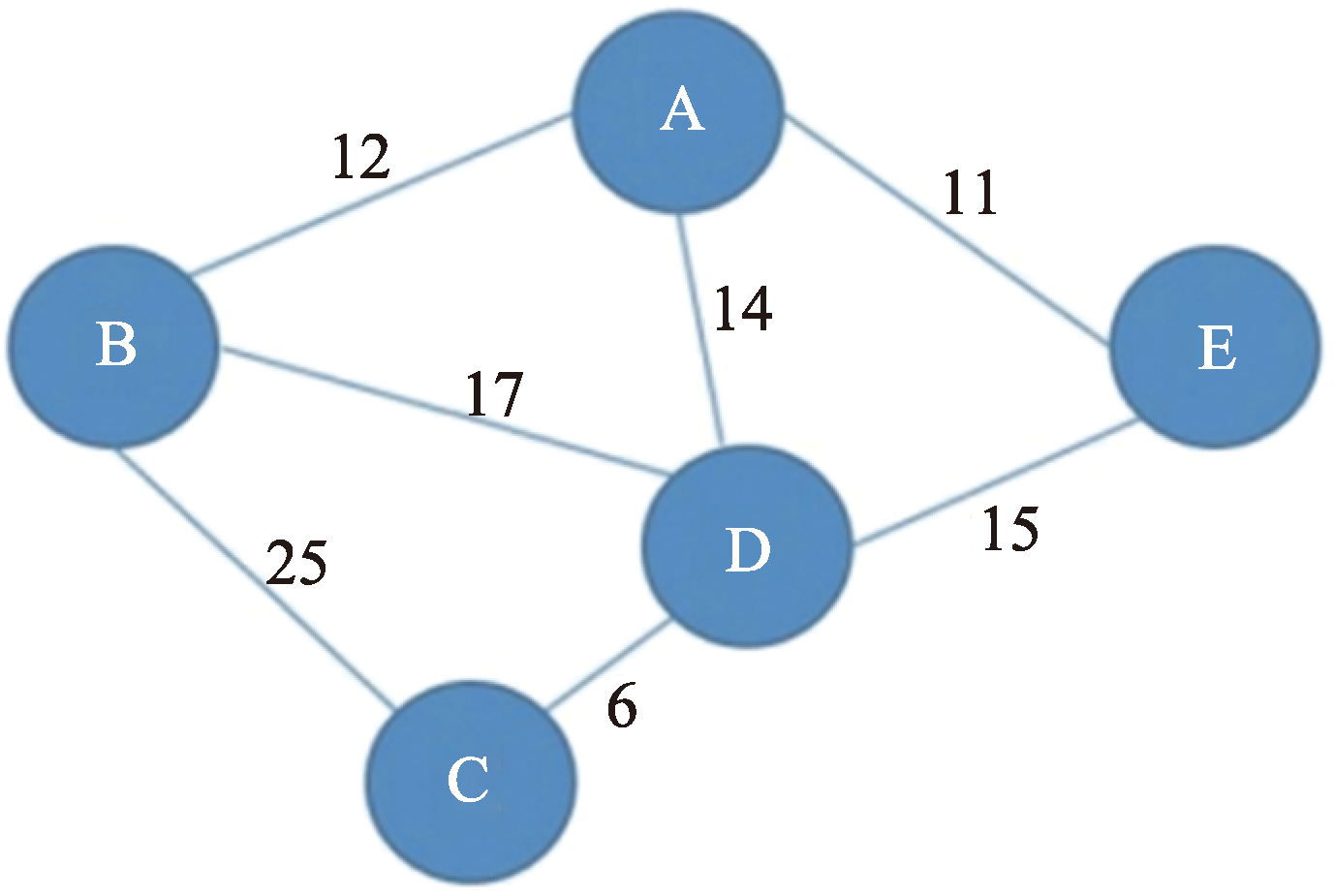
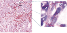
Fig.2
Image construction in a histopathological image (MST algorithm)"

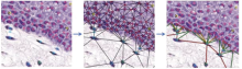
Fig.3
A cell diagram with node and edge markers was constructed using GNN and Delaunay triangulation method[8]"

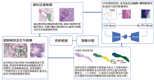
Fig.4
Overview of the application of graph features in image segmentation and detection"

Table 1
Summary of the application of graph method in image segmentation"
| 年份 | 文献 | 技术 | 标签 | 结果评估 |
|---|---|---|---|---|
| 2023 | [ | GNN网络,引入ViG和FPN神经网络模块处理视觉信息 | 有监督 | Dice系数:0.757 |
| 2022 | [ | 先嵌入型残差注意力网络模型 | 有监督 | Dice系数:0.954 |
| 2022 | [ | GraphSegNet网络架构,创新型图卷积网络(GCNN) | 有监督 | Dice系数:0.837 |
| 2021 | [ | SegGini模型 | 弱监督 | Dice系数:0.696 |
Table 2
Summary of application of graph method in object detection"
| 任务 | 年份 | 文献 | 技术 | 结果评估 |
|---|---|---|---|---|
| 细胞/细胞核检测 | 2021 | [ | 构建跨编码成对相似性的不同核组件的空间结构图 | 检测精确度:92.2% |
| 2023 | [ | 基于端到端图的核特征对齐(GNFA)方法 | 检测精确度:76.2% | |
| 2023 | [ | 基于GCN的像素分类器 | 检测精确度:73.4% | |
| 2023 | [ | 基于图的节点分类,利用局部细胞特征和全局组织结构进行上皮细胞检测 | F1-score:0.918 | |
| 病灶区域检测 | 2023 | [ | 基于图的稀疏主成分分析(GS-PCA)网络 | 检测精确度:95.1% |
| 2022 | [ | 基于位置感知图和深度散列(hash)技术,构造位置感知图(LA-Graphs) | 检索精确度:89.5% |
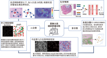
Fig.5
Overview of the application of graph features in image classification"

Table 3
Summary of the application of graph method in image classification"
| 任务 | 年份 | 文献 | 方法 | 标签 | 结果评估 |
|---|---|---|---|---|---|
| 二分类 | 2021 | [ | 结合CNN特征提取和GCN的图像关联学习 | 有监督 | 分类准确度:98.37% |
| 2022 | [ | 复合扩张的主干网络(composite dilated backbone network (CDBN)) | 有监督 | 分类准确度:94.00% | |
| 2023 | [ | CRCCN-Net分类网络模型 | 有监督 | 分类准确度:96.26% | |
| 2023 | [ | 自适应、可扩展的图卷积网络GraphLSurv | 有监督 | concordance-index:0.683,比其他方法提高了3.4% | |
| 2023 | [ | GARL-Net网络,深度神经网络DNN | 有监督 | 分类准确度:99.00% | |
| 2021 | [ | 支持向量机 (SVM) 和 K-Nearest Neighbour (KNN) 算法 | 有监督 | 分类准确度:98.00% | |
| 2022 | [ | GNN+二次谐波生成显微镜图像的胶原纤维形态学特征 | 有监督 | 分类准确度:96.20% | |
| 2023 | [ | 最小化细胞图(Minimized Cellular Graph,MCG) | 有监督 | 分类准确度:97.70% | |
| 2021 | [ | 手工特征(骨骼特征和晶格特征)绘制 | 有监督 | ||
| 2022 | [ | 深度学习模型特征和手工特征组合提取最佳的混合特征 | 有监督 | 分类准确度:91.00% | |
| 2022 | [ | 注意全局上下文图卷积神经网络(AGGCN) | 有监督 | 分类准确度:78.20% | |
| 2022 | [ | 深度特征图注意网络(DeepGAT) | 有监督 | 分类准确度:95.10% | |
| 2021 | [ | 改进对抗生成网络,NAS-SGAN网络 | 半监督 | 分类准确度:98.40% | |
| 2023 | [ | 异构图的框架和异构图边属性转换器HEAT | 伪标签 | 分类准确度:99.10% | |
| 2021 | [ | 基于空间解析的图卷积网络Patch-GCN | 弱监督 | 分类准确度:93.58% | |
| 2021 | [ | 图神经网络(GNN)+ 使用对比损失的自监督训练方法 | 无监督 | 分类准确度:86.00% | |
| 四分类 | 2023 | [ | 分形GCN网络融合到CNN框架中形成CNN-FGCN模型 | 有监督 | 分类准确度:95.77% |
| 五分类 | 2021 | [ | 基于多头注意力的多尺度图网络 | 有监督 | 分类准确度:71.00% |
| 四分类 | 2021 | [ | 高效网的迁移学习方法 | 有监督 | 分类准确度:98.33% |
| 七分类 | 2022 | [ | 图卷积神经网络 | 有监督 | 分类准确度:56.25% |
| 四分类 | 2022 | [ | 集成网络模型CNN-GCN | 有监督 | 分类准确度:94.44% |
| 八分类 | 2022 | [ | 依赖性的轻量级卷积神经网络(DBLCNN) | 有监督 | 分类准确度:99.54% |
| 七分类 | 2022 | [ | 新的层次实体图表示(HACT)和层次学习(HACT-Net)方法 | 有监督 | 分类准确度:83.79% |
| 三分类 | 2022 | [ | 层次变换图神经网络(HAT-Net+) | 无监督 | 分类准确度:98.00% |
| 三分类 | 2022 | [ | 最小生成树MST | 无监督 | 区分低中高密度区,并提取图特征 |
| 三分类 | 2023 | [ | 相关图注意网络(MLP-GAT) | 有监督 | 分类准确度:79.02% |
| [1] | 近30年全球29种癌症的发生率及死亡率[J]. 实用肿瘤学杂志, 2021, 35(06): 561. |
| [2] | MALIK N. R. Graph theory with applications to engineering and computer science[J]. in Proceedings of the IEEE, vol. 63, no. 10, pp. 1533-1534, Oct. 1975.doi: 10.1109/PROC.1975.9996. |
| [3] | CHAKRABORTY M., CHOWDHURY S., CHAKRABO-RTY J, et al. Algorithms for generating all possible spanning trees of a simple undirected connected graph: an extensive review[J/OL]. Complex & Intelligent Systems, 2019, 5: 265-281. https://doi.org/10.1007/s40747-018-0079-7. |
| [4] | ZHOU J, CUI G, HU S, et al. Graph neural networks: A review of methods and applications[J]. AI Open, vol. 1, pp. 57-81, 2020. doi: https://doi.org/10.1016/j.aiopen.2021.01.001. |
| [5] | ZHANG S, TONG H, XU J, et al. Graph convolutional networks: a comprehensive review[J/OL]. Computational Social Networks, 2019, 6, 11. https://doi.org/10.1186/s40649-019-0069-y. |
| [6] |
SCARSELLI F, GORI M, TSOI AC, et al. The graph neural network model[J]. IEEE Transactions on Neural Networks, 2009, 20(1) 61-80.
doi: 10.1109/TNN.2008.2005605 pmid: 19068426 |
| [7] | RUIZ L, GAMA F, RIBEIRO A. Gated Graph Recurrent Neural Networks[J/OL]. in IEEE Transactions on Signal Processing, vol. 68, pp. 6303-6318, 2020.doi: 10.1109/TSP.2020.3033962. |
| [8] | NAIR A, ARVIDSSON H, JORGE V, et al. A graph neural network framework for mapping histological topology in oral mucosal tissue[J]. BMC Bioinformatics, vol. 23, no. 1, Nov. 2022. doi: https://doi.org/10.1186/s12859-022-05063-5. |
| [9] | HAN K, WANG Y, GUO J, et al. Vision GNN: An Image is Worth Graph of Nodes[EB/OL]. Advances in Neural Information Processing Systems, 2022 35: 8291-8303. https://proceedings.neurips.cc/paper_files/paper/2022/file/3743e69c8e47eb2e6d3afaea80e439fb-Paper-Conference.pdf.. |
| [10] | GAO Z, SHI J, WANG J. GQ-GCN: Group quadratic graph convolutional network for classification of histopathological images[C]//Medical Image Computing and Computer Assisted Intervention-MICCAI 2021: 24th International Conference, Strasbourg, France, September 27-October 1, 2021, Proceedings, Part VIII 24. Springer International Publishing, 2021: 121-131. |
| [11] |
ABDLSAMEA M, ZIDAN U, SENOUSY Z, et al. A survey on artificial intelligence in histopathology image analysis[J]. Wiley Interdisciplinary Reviews: Data Mining and Knowledge Discovery, 2022, 12(6): e1474.
doi: 10.1002/widm.v12.6 |
| [12] |
VISWANATHAN V S, TORO P, CORREDOR G, et al. The state of the art for artificial intelligence in lung digital pathology[J]. The Journal of Pathology, 2022, 257(4): 413-429.
doi: 10.1002/path.5966 pmid: 35579955 |
| [13] |
PRABHU S, PRASAD K, ROBELS-KELLY A, et al. AI-based carcinoma detection and classification using histopathological images: A systematic review[J]. Computers in Biology and Medicine, 2022, 142: 105209.
doi: 10.1016/j.compbiomed.2022.105209 |
| [14] |
WU Y, CHENG M, HUANG S, et al. Recent advances of deep learning for computational histopathology: principles and applications[J]. Cancers, 2022, 14(5): 1199.
doi: 10.3390/cancers14051199 |
| [15] |
LINKON A H M, LABIB M M, HASAN T, et al. Deep learning in prostate cancer diagnosis and Gleason grading in histopathology images: An extensive study[J]. Informatics in Medicine Unlocked, 2021, 24: 100582.
doi: 10.1016/j.imu.2021.100582 |
| [16] |
DE MATOS J, ATAKY S T M, DE SOUZA BRITTO J A, et al. Machine learning methods for histopathological image analysis: A review[J]. Electronics, 2021, 10(5): 562.
doi: 10.3390/electronics10050562 |
| [17] | AI S, LI C, LI X, et al. A state-of-the-art review for gastric histopathology image analysis approaches and future development[J/OL]. BioMed Research International, 2021: 6671417. doi: 10.1155/2021/6671417. |
| [18] | LIM S, JUNG S W. A Comparative Study on Graph Construction Methods for Survival Prediction using Histopathology Images[C]// 2022 IEEE International Conference on Consumer Electronics-Asia (ICCE-Asia), IEEE, 2022: 1-4. |
| [19] |
LI M M, HUANG K, ZITNIK M. Graph representation learning in biomedicine and healthcare[J]. Nature Biomedical Engineering, 2022, 6(12): 1353-1369.
doi: 10.1038/s41551-022-00942-x pmid: 36316368 |
| [20] | XIA M, HE Y. Magnetic Resonance Imaging and Graph Theoretical Analysis of Complex Brain Networks in Neuropsychiatric Disorders[J/OL]. Brain Connectivity, vol. 1, no. 5, pp. 349-365, Dec. 2011. doi: https://doi.org/10.1089/brain.2011.0062. |
| [21] | ZHOU Y, YU F, DUONG T. Multiparametric MRI Characterization and Prediction in Autism Spectrum Disorder Using Graph Theory and Machine Learning[J/OL]. PLoS ONE, vol. 9, no. 6, p. e90405, Jun. 2014. doi: https://doi.org/10.1371/journal.pone.0090405. |
| [22] | SHAHABAZ, SOMWANSHI D, YADAV A, et al. Medical images texture analysis: A review[C]. 2017 International Conference on Computer, Communications and Electronics (Comptelix), Jaipur, India, 2017, pp. 436-441. doi: 10.1109/COMPTELIX.2017.8004009 [2017-08-10]. |
| [23] | FAYAZ M, SHAH A, WAHID F, et al. A Robust Technique of Brain MRI Classification using Color Features and K-Nearest Neighbors Algorithm[J/OL]. International Journal of Signal Processing, Image Processing and Pattern Recognition, vol. 9, no. 10, pp. 11-20, Oct. 2016. doi: https://doi.org/10.14257/ijsip.2016.9.10.02. |
| [24] | ASODEKAR B, GORE S, THAKARE A. Brain Tumor analysis Based on Shape Features of MRI using Machine Learning[C]. 2019 5th International Conference On Computing, Communication, Control And Automation (ICCUBEA), Pune, India, 2019, pp. 1-5. doi: 10.1109/ICCUBEA47591.2019.9129512. |
| [25] | HE P, QU A, XIAO S, et al. A GNN-based Network for Tissue Semantic Segmentation in Histopathology Image[C]// Journal of Physics: Conference Series, IOP Publishing, 2023, 2504(1): 012047. |
| [26] |
SHI T, LI C, XU D, et al. Fine-grained histopathological cell segmentation through residual attention with prior embedding[J]. Multimedia Tools and Applications, 2022, 81(5): 6497-6511.
doi: 10.1007/s11042-021-11835-7 |
| [27] | DAMANIA K, ANGEL ARUL J. Graph Convolutional Neural Networks for Nuclei Segmentation from Histopathology Images[C]// International Conference on Soft Computing and its Engineering Applications, Cham: Springer Nature Switzerland, 2022: 158-169. |
| [28] | ANKLIN V, PATI P, JAUME G, et al. Learning whole-slide segmentation from inexact and incomplete labels using tissue graphs[C]//Medical Image Computing and Computer Assisted Intervention-MICCAI 2021: 24th International Conference, Strasbourg, France, September 27-October 1, 2021, Proceedings, Part II 24, Springer International Publishing, 2021: 636-646. |
| [29] |
ZHENG Y, JIANG Z, SHI J, et al. Encoding histopathology whole slide images with location-aware graphs for diagnostically relevant regions retrieval[J]. Medical image analysis, 2022, 76: 102308.
doi: 10.1016/j.media.2021.102308 |
| [30] |
RAM S, TANG W, BELL A, et al. Lung cancer lesion detection in histopathology images using graph-based sparse PCA network[J]. Neoplasia, 2023, 42: 100911.
doi: 10.1016/j.neo.2023.100911 |
| [31] | OZEN Y, AKSOY S, Kösemehmetoğlu K, et al. Self-supervised learning with graph neural networks for region of interest retrieval in histopathology[C]// 2020 25th International conference on pattern recognition (ICPR), IEEE, 2021: 6329-6334. |
| [32] |
JAVED S, MAHMOOD A, DIAS J, et al. Spatially constrained context-aware hierarchical deep correlation filters for nucleus detection in histology images[J]. Medical Image Analysis, 2021, 72: 102104.
doi: 10.1016/j.media.2021.102104 |
| [33] | WANG Z, FAN K, ZHU X, et al. Cross-domain Nuclei Detection in Histopathology Images using Graph-based Nuclei Feature Alignment[J/OL]. IEEE Journal of Biomedical and Health Informatics, 2023. doi: 10.1109/JBHI.2023.3280958. |
| [34] | BAHADE S, EDWARDS M, XIE X. Cascaded Graph Convolution Approach for Nuclei Detection in Histopathology Images[J]. Journal of Image and Graphics, 2023, 11(1). |
| [35] | FREI A, KHAN A, STUDER L, et al. Local and global feature aggregation for accurate epithelial cell classification using graph attention mechanisms in histopathology images[C]// Medical Imaging with Deep Learning, short paper track, 2023[2023-08-04]. |
| [36] |
HASSAN T, JAVED S, MAHMOOD A, et al. Nucleus classification in histology images using message passing network[J]. Medical Image Analysis, 2022, 79: 102480.
doi: 10.1016/j.media.2022.102480 |
| [37] |
SHI J, WANG R, ZHENG Y, et al. Cervical cell classification with graph convolutional network[J]. Computer Methods and Programs in Biomedicine, 2021, 198: 105807.
doi: 10.1016/j.cmpb.2020.105807 |
| [38] | MOHANAKURUP V, PARAMBIL G, GOEL P, et al. Breast cancer detection on histopathological images using a composite dilated Backbone Network[J/OL]. Computational Intelligence and Neuroscience, 2022: 8517706. doi: 10.1155/2022/8517706. |
| [39] |
KUMAR A, VISHWAKARMA A, BAJAJ V. CRCCN-Net: Automated framework for classification of colorectal tissue using histopathological images[J]. Biomedical Signal Processing and Control, 2023, 79: 104172.
doi: 10.1016/j.bspc.2022.104172 |
| [40] |
LIU P, JI L, YE F, et al. GraphLSurv: A scalable survival prediction network with adaptive and sparse structure learning for histopathological whole-slide images[J]. Computer Methods and Programs in Biomedicine, 2023, 231: 107433.
doi: 10.1016/j.cmpb.2023.107433 |
| [41] |
DING S, GAO Z, WANG J, et al. Fractal graph convolutional network with MLP-mixer based multi-path feature fusion for classification of histopathological images[J]. Expert Systems with Applications, 2023, 212: 118793.
doi: 10.1016/j.eswa.2022.118793 |
| [42] |
PATEL V, CHAURASIA V, MAHADEVA R, et al. GARL-Net: Graph Based Adaptive Regularized Learning Deep Network for Breast Cancer Classification[J]. IEEE Access, 2023, 11: 9095-9112.
doi: 10.1109/ACCESS.2023.3239671 |
| [43] | BAKARE Y, KUMARASAMY M. Histopathological image analysis for oral cancer classification by support vector machine[J]. International journal of advances in signal and image sciences, 2021, 7(2): 1-10. |
| [44] |
LI B, NELSON M, SAVARI O, et al. Differentiation of pancreatic ductal adenocarcinoma and chronic pancreatitis using graph neural networks on histopathology and collagen fiber features[J]. Journal of Pathology Informatics, 2022, 13: 100158.
doi: 10.1016/j.jpi.2022.100158 |
| [45] | SAHA P, DAS P, NATH N, et al. Estimation of Abnormal Cell Growth and MCG-Based Discriminative Feature Analysis of Histopathological Breast Images[J/OL]. International Journal of Intelligent Systems, 2023. 10.1155/2023/6318127. |
| [46] | PATI P. JAUME G, FERNANDES L. et al. HACT-Net: A Hierarchical Cell-to-Tissue Graph Neural Network for Histopathological Image Classification[C]. In: Sudre, C.H., et al. Uncertainty for Safe Utilization of Machine Learning in Medical Imaging, and Graphs in Biomedical Image Analysis. UNSURE GRAIL 2020. Lecture Notes in Computer Science, vol 12443. Springer, Cham. https://doi.org/10.1007/978-3-030-60365-6_20. |
| [47] | XIAO R, DEBREUVE E, AMBROSETTI D, et al. Renal cell carcinoma classification from vascular morphology[C]. Medical Image Computing and Computer Assisted Intervention-MICCAI 2021: 24th International Conference, Strasbourg, France, September 27-October 1, 2021, Proceedings, Part VI 24. Springer International Publishing, 2021: 611-621. |
| [48] | SNIGDHA V, NAIR L S. Hybrid feature-based invasive ductal carcinoma classification in breast histopathology images[M]. Machine Learning and Autonomous Systems:Proceedings of ICMLAS 2021, Singapore: Springer Nature Singapore, 2022: 515-525. |
| [49] |
MAHMOOD T, KIM S G, KOO J H, et al. Artificial intelligence-based tissue phenotyping in colorectal cancer histopathology using visual and semantic features aggregation[J]. Mathematics, 2022, 10(11): 1909.
doi: 10.3390/math10111909 |
| [50] | LEE Y, PARK J H, OH S, et al. Derivation of prognostic contextual histopathological features from whole-slide images of tumours via graph deep learning[J]. Nature Biomedical Engineering, 2022: 1-15. |
| [51] | REISENBÜCHLER D, WAGNER S J, BOXBERG M, et al. Local attention graph-based transformer for multi-target genetic alteration prediction[C]// International Conference on Medical Image Computing and Computer-Assisted Intervention, Cham: Springer Nature Switzerland, 2022: 377-386. |
| [52] | ZHANG H, MENG Y, ZHAO Y, et al. DTFD-MIL: Double-tier feature distillation multiple instance learning for histopathology whole slide image classification[C]// Proceedings of the IEEE/CVF Conference on Computer Vision and Pattern Recognition, 2022: 18802-18812. |
| [53] |
BARANWAL M, KRISHNAN S, ONEKA M, et al. Cgat: Cell graph attention network for grading of pancreatic disease histology images[J]. Frontiers in Immunology, 2021, 12: 727610.
doi: 10.3389/fimmu.2021.727610 |
| [54] |
SU R, HE H, SUN C, et al. Prediction of drug-induced hepatotoxicity based on histopathological whole slide images[J]. Methods, 2023, 212: 31-38.
doi: 10.1016/j.ymeth.2023.01.005 pmid: 36706825 |
| [55] | CONG C, YANG Y, LIU S, et al. Imbalanced Histopathology Image Classification Using Deep Feature Graph Attention Network[C]// 2022 IEEE 19th International Symposium on Biomedical Imaging (ISBI), IEEE, 2022: 1-4. |
| [56] |
DAS A, DEVARAMPATI V K, NAIR M S. NAS-SGAN: a semi-supervised generative adversarial network model for atypia scoring of breast cancer histopathological images[J]. IEEE Journal of Biomedical and Health Informatics, 2021, 26(5): 2276-2287.
doi: 10.1109/JBHI.2021.3131103 |
| [57] | SHI J, TANG L, LI Y, et al. A Structure-aware Hierarchical Graph-based Multiple Instance Learning Framework for pT Staging in Histopathological Image[J]. IEEE Transactions on Medical Imaging. vol. 42, no. 10, pp. 3000-3011, Oct. 2023. doi: 10.1109/TMI.2023.3273236. |
| [58] | CHAN T H, CENDRA F J, MA L, et al. Histopathology Whole Slide Image Analysis With Heterogeneous Graph Representation Learning[C]// Proceedings of the IEEE/CVF Conference on Computer Vision and Pattern Recognition, 2023: 15661-15670. |
| [59] | CHEN R J, LU M Y, SHABAN M, et al. Whole slide images are 2d point clouds: Context-aware survival prediction using patch-based graph convolutional networks[C]//Medical Image Computing and Computer Assisted Intervention-MICCAI 2021:24th International Conference, Strasbourg, France, September 27-October 1, 2021, Proceedings, Part VIII 24, Springer International Publishing, 2021: 339-349. |
| [60] |
LEE J, WARNER E, SHAIKHOUNI S, et al. Clustering-based spatial analysis (CluSA) framework through graph neural network for chronic kidney disease prediction using histopathology images[J]. Scientific Reports, 2023, 13(1): 12701.
doi: 10.1038/s41598-023-39591-8 pmid: 37543648 |
| [61] | DIAO S, LUO W, HOU J. Deep Multi-Magnification Similarity Learning for Histopathological Image Classification[J]. IEEE Journal of Biomedical and Health Informatics, vol. 27, no. 3, pp. 1535-1545. Mar. 2023. doi: https://doi.org/10.1109/jbhi.2023.3237137. |
| [62] | DI D, ZOU C, FENG Y, et al. Generating hypergraph-based high-order representations of whole-slide histopathological images for survival prediction[J]. IEEE Transactions on Pattern Analysis and Machine Intelligence, 2022, 45(5): 5800-5815. |
| [63] |
LU C, KOYUNCU C, CORREDOR G, et al. Feature-driven local cell graph (FLocK): new computational pathology-based descriptors for prognosis of lung cancer and HPV status of oropharyngeal cancers[J]. Medical image analysis, 2021, 68: 101903.
doi: 10.1016/j.media.2020.101903 |
| [64] |
SAEDNIA K, LAGREE A, ALERA M A, et al. Quantitative digital histopathology and machine learning to predict pathological complete response to chemotherapy in breast cancer patients using pre-treatment tumor biopsies[J]. Scientific Reports, 2022, 12(1): 9690.
doi: 10.1038/s41598-022-13917-4 pmid: 35690630 |
| [65] |
DING K, ZHOU M, WANG H, et al. Spatially aware graph neural networks and cross-level molecular profile prediction in colon cancer histopathology: a retrospective multi-cohort study[J]. The Lancet Digital Health, 2022, 4(11): e787-e795.
doi: 10.1016/S2589-7500(22)00168-6 |
| [66] |
ALTINI N, PURO E, TACCOGNA M G, et al. Tumor Cellularity Assessment of Breast Histopathological Slides via Instance Segmentation and Pathomic Features Explainability[J]. Bioengineering, 2023, 10(4): 396.
doi: 10.3390/bioengineering10040396 |
| [67] |
HOWARD F M, DOLEZAL J, KOCHANNY S, et al. The impact of site-specific digital histology signatures on deep learning model accuracy and bias[J]. Nature communications, 2021, 12(1): 4423.
doi: 10.1038/s41467-021-24698-1 pmid: 34285218 |
| [68] | XING X, MA Y, JIN L, et al. A Multi-scale Graph Network with Multi-head Attention for Histopathology Image Diagnosis[C]// COMPAY 2021:The third MICCAI workshop on Computational Pathology, 2021, 156: 227-235. |
| [69] | MUNIEN C, VIRIRI S. Classification of hematoxylin and eosin-stained breast cancer histology microscopy images using transfer learning with EfficientNets[J/OL]. Computational Intelligence and Neuroscience, 2021: 5580914. doi: 10.1155/2021/5580914. |
| [70] | TEPE E, BILGIN G. Classification of Tissue Types in Histology Images Using Graph Convolutional Networks[C]// 2022 10th International Symposium on Digital Forensics and Security (ISDFS), IEEE, 2022: 1-3. |
| [71] | DE A, MHATRE R, TIWARI M, et al. Brain Tumor Classification from Radiology and Histopathology using Deep Features and Graph Convolutional Network[C]// 2022 26th International Conference on Pattern Recognition (ICPR), IEEE, 2022: 4420-4426. |
| [72] |
GAO Z, LU Z, WANG J, et al. A convolutional neural network and graph convolutional network based framework for classification of breast histopathological images[J]. IEEE Journal of Biomedical and Health Informatics, 2022, 26(7): 3163-3173.
doi: 10.1109/JBHI.2022.3153671 |
| [73] | DWIVEDI C, NOFALLAH S, POURYAHYA M, et al. Multi stain graph fusion for multimodal integration in pathology[C]// Proceedings of the IEEE/CVF Conference on Computer Vision and Pattern Recognition, 2022: 1835-1845. |
| [74] |
WANG C, GONG W, CHENG J, et al. DBLCNN: Dependency-based lightweight convolutional neural network for multi-classification of breast histopathology images[J]. Biomedical Signal Processing and Control, 2022, 73: 103451.
doi: 10.1016/j.bspc.2021.103451 |
| [75] | CHEN H, WANG K, ZHU Y, et al. From pixel to whole slide: Automatic detection of microvascular invasion in hepatocellular carcinoma on histopathological image via cascaded networks[C]//Medical Image Computing and Computer Assisted Intervention-MICCAI 2021: 24th International Conference, Strasbourg, France, September 27-October 1, 2021, Proceedings, Part VIII 24, Springer International Publishing, 2021: 196-205. |
| [76] |
PATI P, JAUME G, FONCUBIERTA-RODRIGUEZ A, et al. Hierarchical graph representations in digital pathology[J]. Medical image analysis, 2022, 75: 102264.
doi: 10.1016/j.media.2021.102264 |
| [77] |
BAI Y, MI Y, SU Y, et al. A scalable graph-based framework for multi-organ histology image classification[J]. IEEE Journal of Biomedical and Health Informatics, 2022, 26(11): 5506-5517.
doi: 10.1109/JBHI.2022.3199110 |
| [78] |
PEREGRINA-BARRETO H, RAMIREZ-GUATEMALA V Y, LOPEZ-ARMAS G C, et al. Characterization of Nuclear Pleomorphism and Tubules in Histopathological Images of Breast Cancer[J]. Sensors, 2022, 22(15): 5649.
doi: 10.3390/s22155649 |
| [79] | YANG X, SHANG L, HE R, et al. Clustering of Cervical Histopathology Images Based on Minimum Spanning Tree[C]// International Conference on Image, Vision and Intelligent Systems. Singapore: Springer Nature Singapore, 2022: 92-101. |
| [80] | LIU L, WANG Y, CHANG J, et al. A correlation graph attention network for classifying chromosomal instabilities from histopathology whole-slide images[J]. iScience, 2023, 26(6).[2022-11-15]. |
| [81] | REN J, RAJBHANDARI S, AMINABADI R Y, et al. ZeRO-Offload: Democratizing Billion-Scale model training[C]// 2021 USENIX Annual Technical Conference (USENIX ATC 21), 2021: 551-564. |
| [82] | RASLEY J, RAjBHANDARI S, RUWASE O, et al. Deepspeed: System optimizations enables training deep learning models with over 1000an parameters[C]// Proceedings of the 26th ACM SIGKDD International Conference on Knowledge Discovery & Data Mining, 2020: 3505-3506. |
| [1] | YE Xu, DU Yi, CUI Wenjuan, SHEN Junjie, XIE Jing, WANG Ludi. Application of Machine Learning Technology in the Field of Eye Health [J]. Frontiers of Data and Computing, 2024, 6(2): 117-133. |
| [2] | SHEN Zhihao, LI Na, YIN Shihao, DU Yi, HU Lianglin. Airfare Price Prediction Based on TPA-Transformer [J]. Frontiers of Data and Computing, 2023, 5(6): 115-125. |
| [3] | WEI Ting, PENG Liang, NIU Tie, ZHANG Honghai. Detection and Root Cause Analysis of HPC Failure Jobs Based on Feature Analysis [J]. Frontiers of Data and Computing, 2023, 5(6): 94-103. |
| [4] | SUN Yifan, ZHANG Rui, TAO Yang, GAO Birou, QIN Shihan, AN Chao. A Survey on Local Differential Privacy [J]. Frontiers of Data and Computing, 2023, 5(5): 74-97. |
| [5] | TIAN Yiqing, CHENG Xi, FENG Bojing. A Review of Computational Models for Corporate Credit Rating [J]. Frontiers of Data and Computing, 2023, 5(4): 139-153. |
| [6] | CHEN Meilin, LIU Duanyang, XU Liming, WANG Yang. A Review of Force Field Models Based on Machine Learning [J]. Frontiers of Data and Computing, 2023, 5(4): 27-37. |
| [7] | LIU Duanyang, WEI Zhongming. Application of Supervised Learning Algorithms in Materials Science [J]. Frontiers of Data and Computing, 2023, 5(4): 38-47. |
| [8] | ZHU Mingming, CAO Wudi, WU Lin, WANG Zixi, LIAO Qi, ZHANG Si, TANG Xiao, LI Jie, WANG Jing, WANG Yangang, WANG Zifa. The Development and Prospects of Informatization Applications in Dual-Carbon Atmospheric Environment Based on Artificial Intelligence and Big Data [J]. Frontiers of Data and Computing, 2023, 5(3): 2-12. |
| [9] | LI Yan,HE Hongbo,WANG Runqiang. A Survey of Research on Microblog Popularity Prediction [J]. Frontiers of Data and Computing, 2023, 5(2): 119-135. |
| [10] | HU Xiaoyan,XU Jiyao,ZOU Ziming. Preliminary Study on Paradigm Shift in Space Weather Research Driven by Big Data and Artificial Intelligence [J]. Frontiers of Data and Computing, 2023, 5(2): 24-36. |
| [11] | QI Fazhi,LI Gang,LI Chun,WANG Lu,ZHANG Yi,ZHANG Zhengde,CHEN Gang,LUO Wuming,ZHAO Lina,HU Yu,YUAN Ye. Big Data and AI for High Energy Physics [J]. Frontiers of Data and Computing, 2023, 5(2): 50-59. |
| [12] | GAO Tian,ZHU Jiaojun,ZHANG Jinxin,SUN Yirong,YU Fengyuan,TENG Dexiong,LU Deliang,YU Lizhong,WANG Zongguo. Estimation of Carbon Flux of a Temperate Forest Ecosystem Based on Next-Generation Information Technologies [J]. Frontiers of Data and Computing, 2023, 5(2): 60-72. |
| [13] | WANG Fan,FENG Liqiang,CAO Rongqiang. Design and Application of Big Data-Driven Ocean Artificial Intelligence Service Platform [J]. Frontiers of Data and Computing, 2023, 5(2): 73-85. |
| [14] | WANG Zongguo,WAN Meng,CHEN Ziyi,LI Kai,WANG Xiaoguang,LIU Miao,MENG Sheng,WANG Yangang. Research and Application of a Data-Driven Intelligent Design Platform for Materials [J]. Frontiers of Data and Computing, 2023, 5(2): 86-96. |
| [15] | LIU Jiaqi,YANG Binyan. Research on the Hot Spots and Trends of the Coupling Development of Artificial Intelligence and Social Science in China——A Bibliometric Analysis Based on CiteSpace [J]. Frontiers of Data and Computing, 2022, 4(6): 77-91. |
| Viewed | ||||||
|
Full text |
|
|||||
|
Abstract |
|
|||||